Author : Suraj Venna, MD, FAAD Director, Melanoma Center Washington Cancer Institute 110 Irving Street, NW Washington DC 20010 (o) 202 877 2551
2009-07-20
2009-07-20
Actinic Keratoses : pre cancerous skin lesions
Contents
Introduction
What are actinic keratoses and how are they related to skin cancer?
What do actinic keratoses look like?
Who gets actinic keratoses and what are the risk factors?
Is a biopsy needed to make a diagnosis of an actinic keratosis?
How are actinic keratoses treated?
Is there a way to prevent the onset of actinic keratoses?
Introduction
There are two basic categories of skin cancer for which people seek the care of a dermatologist: melanoma and non-melanoma skin cancer (NMSC).
There are 55,000 cases of melanoma annually in the United States and
nearly 1,000,000 cases of NMSC. NMSC includes basal cell skin cancer,
accounting for 80% of cases, and squamous cell skin cancer, accounting
for 20% of cases. Melanoma skin cancer is the most dangerous, since it
appears to have a greater ability to metastasize or spread from the skin
to other body sites. NMSCs have a much lower risk of spreading from the
skin to other body sites. Of the NMSCs, basal cell rarely spreads
beyond the skin, however squamous cell does have a small but real risk
of spread. In the United States, of the nearly 200,000 cases of squamous
cell skin cancer annually, about 1% or 2000 people, die from metastatic
squamous cell skin cancer.
This article examines actinic keratoses (AKs), which are pre-cancerous growths
that can convert to squamous cell skin cancer in as many as 10% of the
people who have AKs.
What are actinic keratoses and how are they related to skin cancer?
Actinic
keratoses (AKs), also known as Solar Keratoses, are one of the most
common reasons for a person to see their dermatologist. AKs are precursor
lesions to the squamous cell type of skin cancer and are considered
pre-cancerous growths. AKs most commonly occur on sun exposed sites of
fair skinned individuals. Although it is traditionally taught that 40%
of squamous cell skin cancers (SCCs) begin as an AK, various studies
over the last 20 years have determined a conversion rate of AKs to SCC
to be 0.1% to 10% (1-3). The vast majority of patients who develop SCC
are effectively treated. A small percentage of these patients, however,
develop life-threatening spread beyond the skin.
AKs
may resolve spontaneously, especially in those who use sunblock and
limit sun exposure. Otherwise AKs may grow into SCCs. Having numerous
AKs are indicative of excessive sun exposure. As such, AKs are markers
for the development of all types of skin cancer, including melanoma and
basal cell type (4-8).
What do actinic keratoses look like?
AKs
are red, scaly growths or bumps, generally smaller than the eraser on a
pencil, with a characteristic gritty, rough or prickly surface. As they
are caused by sun exposure, they commonly occur on the scalp, ears,
face, lips, neck, forearms and the backs of the hands (Figure 1 and
Figure 2). Aside from their appearance, AKs are often asymptomatic, but
they can become thick, bleed, and cause irritation or pain (Figure 3).

Figure 1
Multiple rough lesions on the face and scalp of an elderly person.
Also note AKs on the rim of the ear.

Figure 2
Sun exposed area of the hand showing multiple actinic keratoses.

Untreated, diffuse sun damage with thick actinic keratoses.
Who gets actinic keratoses and what are the risk factors?
About
10-20% of adults in the United States have AKs. These lesions are
primarily caused by ultraviolet radiation from the sun and, if left
untreated, can transform into SCC (Figure 4). AKs occur predominantly in
the Caucasian population and correlate with the amount of sun exposure
over one’s lifetime. People usually begin to develop AKs in their
forties and fifties, although AKs may also be seen in people in their
twenties, which may reflect the fact that nearly 80% of sun damage to
the skin has accumulated by the time an individual is 18 years old. AKs
are more common in men than women and are more likely to occur on
someone who has an outdoor occupation. The highest frequency of AKs
occurs in populations closest to the equator, since these areas are
subjected to more direct and intense sunlight. The highest incidence of
skin cancer in the world is in Australia where nearly 40% of adults have
AKs.
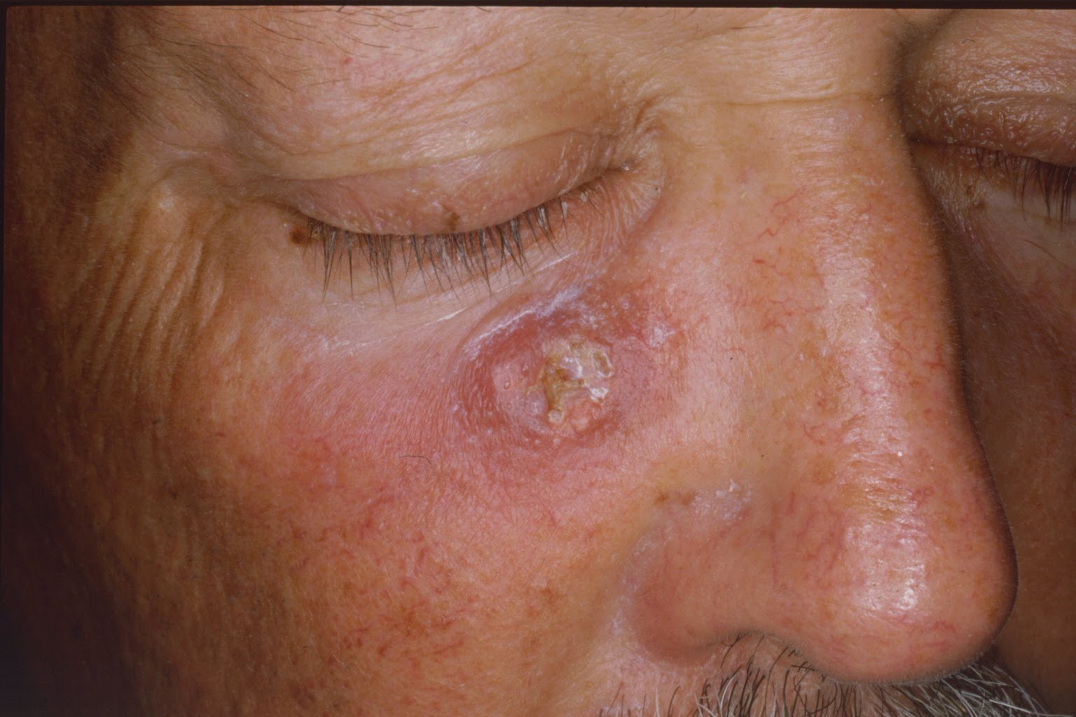
Figure 4
A large squamous cell carcinoma on the cheek.
People
who tend to burn and have a minimal tanning response, also known as
Fitzpatrick skin-type I and II, generally are more susceptible to AKs
and skin cancer compared to darker complexioned people. The reason for
this difference is based on the function of pigmentation in the skin.
Skin pigment acts as a layer of protection against the harmful rays of
the sun. Due to this protective role of pigment, skin cancer and AKs are
rare in darker skinned people. The ability of our bodies to combat
cancer is also a function of our age and the robustness of our immune
system. Thus, individuals who are immunosuppressed are at higher risk of
AKs and skin cancer. Examples of immunosuppressed states include the
following: people taking medications for cancer or autoimmune diseases,
organ transplant recipients, and individuals with acquired
immunodeficiency syndrome (AIDS).
Is a biopsy needed to make a diagnosis of an AK?
The
majority of AKs are diagnosed clinically (through an observation by a
physician); very few are confirmed with a biopsy. The diagnostic
accuracy for clinically identifying an AK correctly is in the range of
74-94% (5,10,11). Nevertheless, when the growth is large, grows rapidly,
causes pain, or has been treated multiple times and is not resolving it
would be prudent to biopsy growths to rule-out skin cancer.
Most
physicians can diagnose an actinic keratosis, although the diagnosis is
most frequently made by dermatologists. Of the 127 million office
visits to dermatologists over a four-year period, 14.6 million or 11.5%
were for the evaluation and treatment of AKs (9).
How are actinic keratoses treated?
It
is important to realize that AKs can be completely and effectively
treated in a variety of ways. Since AKs are readily identified by
physicians, biopsies are not routinely performed and the physician
simply proceeds with treatment. The type of treatment that your
physician may advocate will depend on the location of these lesions, the
number of AKs, the size of the AKs, as well as the medical history of
the patient and age. Most therapies are well tolerated. They can be
broadly divided into surgical and medical treatments as seen in the
table below. The most common treatment is with cryosurgery, followed by
medicated creams and in some instances a combination approach.
| Medical | Surgical |
| Topical chemotherapy | Cryosurgery |
| Topical immunomodulators | Laser resurfacing |
| Chemical peel | Dermabrasion |
| Topical nonsteroidal anti-inflammatory | Curettage |
| Photodynamic therapy | Excisional removal |
Cryosurgery
The most common treatment for AKs is cryosurgery using liquid nitrogen (LN2) at a temperature of about -195 degrees celsius. The LN2
is applied either with a Q-tip, metal surgical instrument or, most
commonly, with an insulated spray thermos. Usually a lesion is treated
twice, with each freezing cycle lasting a few seconds. A transient
stinging sensation develops, after which the area becomes red and may
blister. Over the course of days to weeks, the lesion will crust over
and scale off or at least will decrease in size. Some patients will
develop whitish scars at treated sites, which in some instances can be
permanent. The use of LN2 is best suited for those who have
less than 10-15 lesions. Beyond this number of lesions, treatments that
can elicit more of a field effect are used. Generally, AKs should revert
or disappear after 2 treatments (each treatment consisting of two
rounds of LN2). If a lesion continues to grow in spite of
treatment, it is prudent to have this lesion biopsied or sampled,
because it may have transformed into squamous cell skin cancer or may be
something other than an AK or SCC (5).
Medicated creams
In
patients with many AKs, a medicated cream may be the best approach
since this will allow treatment of a greater area and cover many lesions
(>10 -15). The other benefit to topical treatment is their ability
to halt the aberrant growth of single, atypical cells in the skin that
constitute an AK, even though they don’t always form an obvious growth
or lesion. However, these single or few atypical cells will succumb to
the effects of topical treatment; this is known as the field effect.
Topical chemotherapy: fluorouracil
Atypical
cells, as found in AKs, divide faster than normal healthy skin cells.
This is the basis for the action of fluorouracil: it becomes
incorporated into cells that are dividing rapidly and once in the cell,
prevents the cell from further divisions, ultimately eradicating this
pre-cancer growth. Fluorouracil is a highly effective, FDA approved
treatment of AKs and has been in use for several decades. Most
formulations are used once or twice per day for four to six weeks and
the patient is educated on the different phases that the skin will
endure. During weeks one and two, there will be irritation, redness,
stinging, crusting and burning. By week three, most patients will go
through an exfoliation phase during which most of the AKs and
damaged skin flakes off and new healthy skin cells replenish the treated
areas. Most patients also notice redness and inflammation on parts of
the skin where they have no obvious AK lesion. This is because of the
aforementioned field effect, where fluorouracil becomes
incorporated into unhealthy skin cells that have not yet formed a lesion
or growth. Patients should be forewarned that they will have a
significant response especially during the first couple of weeks of
application.
Topical immunotherapy: imiquimod
Imiquimod
is a cream that was initially developed and approved for the treatment
of genital warts. It was subsequently approved by the FDA for treatment
of AKs in 2004. This cream stimulates the local immune system in the
skin to destroy the atypical cells forming AKs. As with other topicals,
patients will experience redness, burning and scaling. A small
percentage of patients can also develop flu-like symptoms. The duration
of treatment is up to 16 weeks and patients should apply the cream 2 or 3
times per week. Compared to fluorouracil, the response to imiquimod is
not as predictable: some patients have intense redness and scaling while
others have minimal or no response. This may be a function of the
individual patient’s immune system.
Topical nonsteroidal anti-inflammatory (NSAIDS)
NSAIDs
are a familiar class of medications, examples of which are ibuprofen
(Advil, Motrin) and naproxen (Alleve, Naprosyn). Diclofenac gel is a
topical NSAID approved for the treatment of AKs. The mechanism of action
is not entirely clear, but may in part be related to the medication
blocking nutrients that atypical cells require to grow. Diclofenac gel
is applied twice daily for 60-90 days and, as with other topicals, the
most common side effect is local irritation. People allergic to the
various pill forms of NSAIDs should not use diclofenac.
Photodynamic therapy (PDT)
A
light-sensitive topical solution is applied to the affected area of
skin and gets absorbed into the atypical skin cells. The chemical
applied is called 5-aminolevulinic acid (5-ALA), a compound found in
red-blood cells that is a precursor to hemoglobin (the oxygen carrying
component of blood). After a prescribed time period (few hours to
overnight), the skin is exposed to a special light that activates 5-ALA,
ultimately destroying the actinic keratosis. PDT is thought to offer
more selective destruction over other topical modalities with less
damage to surrounding normal skin. Most patients can see a dramatic
improvement even after one treatment.
The other types of surgical and medical treatments in the above table are not used as commonly, but offer both physicians and patients a wide array of options. Curettage involves the use of a special instrument, the curette, to scrape off the AK, which can then be sent for biopsy evaluation. This is effective for thicker AKs and is often followed by cautery which stops bleeding that may be associated with AK removal. Excision involves numbing the area with local anesthesia then taking a blade and cutting the lesion out, repairing the skin with sutures. The specimen is then sent for biopsy evaluation. This technique will offer complete removal of the lesion, although will leave a scar and is reserved for AKs that are thick and unresponsive to LN2, or when the physician believes the lesion may have already turned into squamous cell skin cancer. Chemical peels, dermabrasion, and lasers are also used for the treatment of AKs. These approaches are based on superficial and deeper skin destruction with removal of the top layer of skin, the epidermis, and sometimes the next layer of skin called the dermis. Since AKs are atypical cells that grow within the epidermis, these resurfacing approaches can be effective, although they are not routinely used for this purpose.
Is there a way to prevent the onset of actinic keratoses?
Since AKs are sun-induced growths, limiting sun exposure is a way to minimize the number of AKs that one gets. A study in the New England Journal of Medicine
found that regular sunscreen use prevents the development of AKs,
reduces the number of existing AKs, and is associated with a potential
risk reduction in skin cancer development (11).
An
intriguing study examined the effects of a low-fat diet on the
subsequent incidence of AKs in a population of patients who have had
skin cancer (12). These people were followed for a two year period: one
group had a diet consisting of 40% fat calories while the intervention
group was restricted to 20% fat calories. Over the two year period, the
patients on the lower fat diet developed on average three AKs compared
to 10 AKs in the unrestricted group. This is an interesting study, but
limited due to the overall number of patients and warrants further
validation.
The key, then, to prevention
is to limit unnecessary and dangerous sun exposure by protecting your
skin through the use of protective sun clothing and sunscreens, avoiding
sunburns, and limiting outdoor activity during peak sun hours
(10am-2pm).
For more information:
http://www.aad.org/media/background/factsheets/fact_sunscreen.htm
References
1. Marks
R, Foley P, Goodman G, et al. Spontaneous remission of solar keratoses:
the case for conservative management. Br J Dermatology 1986; 115:69-55
2. Marks
R, Rennie G, Selwood TS. Malignant transformation of solar keratoses to
squamous cell carcinoma in the skin: a prospective study. Lancet 1988;
795-7
3. Dobson JM, DeSpain J, Hewett JE, Clark DP. Malignant
potential of actinic keratoses and the controversy over treatment: a
patient oriented perspective. Archives of Dermatol. 1991; 127: 1029-31
4. Green
A, Battistutta D. Incidence and determinants of skin cancer in a high
risk Australian population. Int J Cancer 1990; 15:356-61
5. Marks
R, Rennie G, Selwood TS. The relationship of basal cell carcinoma and
squamous cell carcinoma to solar keratoses. Archives of Dermatology
1988; 1124:1039-42
6. Bataille
V, Sasieni P, Grulich A, et al. Solar Keratoses: A Risk Factor for
Melanoma but Negative Association with Melanocytic Nevi. Int J Cancer
1998; 78:8-12
7. Green AC, O’Rourke MG. Cutaneous melanoma in association with other skin cancers. J Nat. Cancer Inst. 1985; 74:977-980
8. Dubin
M, Moseson M, Pasternak BS. Epidemiology of malignant melanoma:
pigmentary traits, ultraviolet radiation and the identification of
high-risk populations. Recent Results Cancer Reasearch 1986;102:56-75
9. Feldman
S, Fleischer AB, McConnell C. Most common dermatologic problems
identified by internists: 1990-94; Archive of Internal Med 1998;
158:726-30
10. Venna
S, Lee D, Stadecker M, Rogers G. Clinical Recognition of Actinic
Keratoses: How Good Are We? Archive of Dermatol 2005;141:507-09
11. Thompson S, Jolley D, Marks R. Reduction of Solar Keratoses by Regular Susncreen Use. NEJM 1993;329:1147-1151
12. Black H, Herd A, Goldberg L et al. Effect of a Low-Fat Diet on the Incidence of Actinic Keratoses. NEJM 1994;330:1272-75